Human spines are complex. No one appreciates that more than the specialists who bring their skills to bear on the complex mass of interwoven bones, nerves, muscles, tendons, and ligaments during a back operation.

That’s why specialists at Braemar Hospital use state-of-the art technology that will make their job safer, easier and provide greater surety for patients.

The O-Arm surgical imaging and guidance system takes pictures and provides an inside view of what’s happening with the spine before and during surgery. It is different from a traditional X-ray machine or CT scanner because it can provide “real-time” and high quality images in two or three dimensions and a detailed look at the anatomy during an operation.

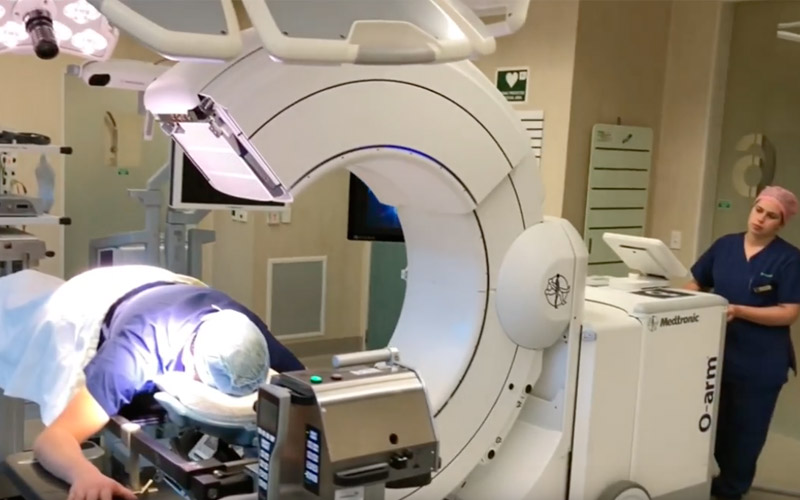
It has been likened to the difference between tracking a geographic location with a paper map and having GPS in your car or mobile device. Braemar Hospital bought the device in 2016 and was the first hospital in the region to use the machine. The technology is also used at both of New Zealand’s spinal trauma centres in Auckland and Christchurch.

Braemar Hospital CEO Fiona Michel says consistently investing in new equipment such as the O-Arm is what sets Braemar apart. “We’re all about making lives better, and while that means caring for our patients, we are excited to be able to provide high-end equipment such as this for our specialists.”

The O-Arm looks like a giant letter ‘O’, hence the name. But the technology behind it is complex. It gives surgeons the ability to navigate a 3-D map of the body part undergoing surgery, which allows them to pinpoint more accurately the exact position of surgical instruments during a procedure. Surgeons say it’s like a “second-sight.”

The improved technology reduces invasiveness and risk for patients; it also allows surgeons to make informed decisions during surgery and precisely navigate through delicate anatomy avoiding critical structures in the spine. Because of the heightened level of accuracy, there is less chance a procedure will need to be revisited.

While the image quality is similar to a CT image, radiation exposure is significantly reduced, making the procedure safer. The machine needs a special table to sit on and this table was paid for by the Braemar Charitable Trust, the hospital’s owner.

“Surgical equipment has been a rapidly changing area and at Braemar we feel it’s vital to keep pace with surgical innovation so we can deliver the best-possible environment for our specialists and our patients.”

Meanwhile Braemar Hospital was the first hospital in Australasia to install ultra-high definition (4K) imaging in its operating theatres, enabling surgeons to carry out laparoscopic (keyhole) surgery with images that closely replicate what their naked eye would see looking into the body.


March 2024